Entradas

Mostrando las entradas de 2018

Estimating the prevalence at death of CTE neuropathology among professional football players

Imagen
In July 2017,   JAMA   published results from autopsies of the brains of 202 deceased American football players. 1   Of the 111 participants who had played in the National Football League (NFL), 110 (99%) received a neuropathologic diagnosis of chronic traumatic encephalopathy (CTE), a progressive neurodegenerative disease associated with head trauma. This 99% figure received substantial media coverage when the study was published. REFERENCE; Binney ZO, Bachynski KE. Estimating the prevalence at death of CTE  neuropathology among professional football players. Neurology. 2018 Nov 28. pii:   10.1212/WNL.0000000000006699. doi: 10.1212/WNL.0000000000006699. [Epub ahead of  print] PubMed PMID: 30487144. Commented on: InsideScience: What's the Risk of Chronic Traumatic Encephalopathy for NFL Players? New study suggests at least one in 10 NFL players could eventually develop the brain disease.

TWiV 453: Neurovirology with Diane Griffin

From the Vector-Borne Viruses Symposium in Hamilton, Montana, Dickson and Vincent speak with Diane Griffin about her career and her work on understanding viral infections of the central nervous system. Hosts: Vincent Racaniello and Dickson Despommier Guest: Diane Griffin TWiV 453 (39 MB .mp3, 64 min) LINKS FOR THIS EPISODE Vector-Borne Viruses Symposium Diane Griffin Adoptive immunization of immunosuppressed mice infected with Sindbis virus Role of the immune response in age-dependent resistance of mice to encephalitis due to Sindbis virus Antibody-mediated clearance of alphavirus infection from neurons Persistence of viral RNA in mouse brains after recovery from acute alphavirus encephalitis Conversion of lytic to persistent alphavirus infection by the bcl-2 cellular oncogene Bc1-2 protects mice against fatal alphavirus encephalitis

8º Curso de Animales de Laboratorio #INR 12-14/SEP/2018

 ·

A "Driver Switchover" Mechanism of Influenza Virus Transport from Microfilaments to Microtubules.

Imagen
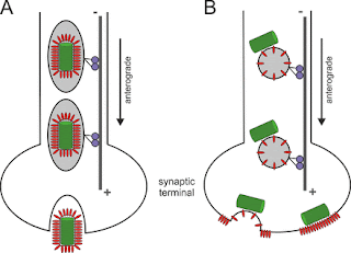
When infecting host cells, influenza virus must move on microfilaments (MFs) at the cell periphery and then move along microtubules (MTs) through the cytosol to reach the perinuclear region for genome release. But how viruses switch from the actin roadway to the microtubule highway remains obscure. To settle this issue, we systematically dissected the role of related motor proteins in the transport of influenza virus between cytoskeletal filaments in situ and in real-time using quantum dot (QD)-based single-virus tracking (SVT) and multicolor imaging. We found that the switch between MF- and MT-based retrograde motor proteins, myosin VI (myoVI) and dynein, was responsible for the seamless transport of viruses from MFs to MTs during their infection. After virus entry by endocytosis, both the two types of motor proteins are attached to virus-carrying vesicles. MyoVI drives the viruses on MFs with dynein on the virus-carrying vesicle hitchhiking. After role exchanges at actin-microtubul

Inhibition of Retrograde Transport Limits Polyomavirus Infection In Vivo

Imagen
Polyomaviruses (PyVs) silently infect most humans, but they can cause life-threatening diseases in immunocompromised individuals. The JC polyomavirus (JCPyV) induces progressive multifocal leukoencephalopathy, a severe demyelinating disease in multiple sclerosis patients receiving immunomodulatory therapy, and BK polyomavirus (BKPyV)-associated nephropathy is a major cause of kidney allograft failure. No effective anti-PyV agents are available. Several compounds have been reported to possess anti-PyV activity in vitro, but none have shown efficacy in clinical trials. Productive PyV infection involves usurping the cellular retrograde vesicular transport pathway to enable endocytosed virions to navigate to the endoplasmic reticulum where virion uncoating begins. Compounds inhibiting this pathway have been shown to reduce infection by simian virus 40 (SV40), JCPyV, and BKPyV in tissue culture. In this study, we investigated the potential of Retro-2.1, a retrograde transport inhibitor, t

Anterograde transport of rabies virus in DRG neurons.

Imagen
Model of intraneuronal postreplicative RABV transport. Rabies  virus  (RABV) spread is widely accepted to occur only by  retrograde  axonal  transport . However, examples of anterograde RABV spread in peripheral  neurons  such as dorsal root ganglion (DRG)  neurons  indicated a possible bidirectional  transport  by an uncharacterized mechanism. Here, we analyzed the axonal  transport  of fluorescence-labeled RABV in DRG  neurons  by live-cell microscopy. Both entry-related  retrograde   transport  of RABV after infection at axon endings and postreplicative  transport  of newly formed  virus  were visualized in compartmentalized DRG neuron cultures. Whereas entry-related  transport  at 1.5 μm/s occurred only retrogradely, after 2 days of infection, multiple particles were observed in axons moving in both the anterograde and  retrograde  directions. The dynamics of postreplicative  retrograde   transport  (1.6 μm/s) were similar to those of entry-related  retrograde   transport . In

Flaviviruses are neurotropic, but how do they invade the CNS?

Imagen
Flaviruses (FV) are RNA viruses carried by mosquitoes. Neurological signs including acute encephalitis, meningitis and acute flaccid paralysis develop in a small percentage of infected individuals; long term sequlae are, Parkinsonism, dystonias and cognitive changes. FV neuroinfection is neurotropic involving subcortical nuclei (substantia nigra and thalamus) anterior horn neurons and neocortex. Glycosylation of the FV E envelope protein is one determinant of neuroinvasion, increasing both axonal and trans-epithelial transportation. Neutralizing antibodies against the E and NS proteins prevents FV uptake into several cell types, including axons. CD8+ T cells are vital for clearance of WNF infected cells from the CNS, whereas TLR-3 and TLR-7 mediated anti-virus response through increased serum inflammatory cytokines to disrupt the BBB providing infected leucocytes and free virus access to the CNS (so called Trojan horse) Cellular virus attachment factors, promoting FV cell entry are w

Prions, prionoids and protein misfolding disorders #PMD

Imagen
Prion diseases are progressive, incurable and fatal neurodegenerative conditions. The term ‘prion’ was first nominated to express the revolutionary concept that a protein could be infectious. We now know that prions consist of PrPSc, the pathological aggregated form of the cellular prion protein PrPC. Over the years, the term has been semantically broadened to describe aggregates irrespective of their infectivity, and the prion concept is now being applied, perhaps overenthusiastically, to all neurodegenerative diseases that involve protein aggregation. Indeed, recent studies suggest that prion diseases (PrDs) and protein misfolding disorders (PMDs) share some common disease mechanisms, which could have implications for potential treatments. Nevertheless, the transmissibility of bona fide prions is unique, and PrDs should be considered as distinct from other PMDs. REFERENCE: Scheckel C, Aguzzi A. Prions, prionoids and protein misfolding disorders. Nat Rev Genet. 2018 Jul;19(7):

Clinical Trials in Neurovirology: Successes, Challenges, and Pitfalls

Imagen
Clinical trials in neurovirology illustrate the special challenges confronting investigators planning to study these conditions, as well as the contributions of successful trials in establishing appropriate management for these devastating diseases. This article reviews key examples of progress in neurovirology that have been spurred by clinical trials, emphasizing human herpes virus encephalitis, HIV, and JC virus. Clinical trials in the setting of neurovirological diseases are characterized by specific challenges, which may include small sample sizes, clinical presentations from life-threatening conditions to chronic courses of disease, regional and temporally restricted outbreaks scenarios, and the unavailability of validated diagnostic tests that can be rapidly deployed at the bedside. This review aims to highlight these methodological challenges and pitfalls in designing and executing clinical neurovirology trials, as well as to outline innovative trial designs, which could be

#brain: The mysterious origins of microglia

Imagen
The origin of microglia, the resident macrophage population of the CNS, has been a long-standing matter of debate. Here we discuss two seminal studies published in 2007 in Nature Neuroscience that significantly contributed to a better understanding of microglia ontogeny and homeostasis in the adult brain. REFERENCE: Ginhoux F, Garel S. The mysterious origins of microglia. Nat. Neurosci. 2018 Jul;21(7):897-899. doi: 10.1038/s41593-018-0176-3.

Viral Triggers and Inflammatory Mechanisms in Pediatric Epilepsy. Review

Imagen
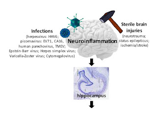
Experimental and clinical findings suggest a crucial role for inflammation in the onset of pediatric seizures; this mechanism is not targeted by conventional antiepileptic drugs and may contribute to refractory epilepsy. Several triggers, including infection with neurotropic viruses such as human herpesvirus 6 (HHV-6), other herpesviruses, and picornaviruses, appear to induce activation of the innate and adaptive immune systems, which results in several neuroinflammatory responses, leading to enhanced neuronal excitability, and ultimately contributing to epileptogenesis. This review discusses the proposed mechanisms by which infection with herpesviruses, and particularly with HHV-6, and ensuing inflammation may lead to seizure generation, and later development of epilepsy. We also examine the evidence that links herpesvirus and picornavirus infections with acute seizures and chronic forms of epilepsy. Understanding the mechanisms by which specific viruses may trigger a cascade of alt

Differential antiviral immunity to Japanese encephalitis virus in developing cortical organoids

Imagen
Japanese encephalitis (JE) caused by Japanese encephalitis virus (JEV) poses a serious threat to the world's public health yet without a cure. Certain JEV-infected neural cells express a subset of previously identified intrinsic antiviral interferon stimulated genes (ISGs), indicating brain cells retain autonomous antiviral immunity. However, whether this happens in composited brain remains unclear. Human pluripotent stem cell (hPSC)-derived organoids can model disorders caused by human endemic pathogens such as Zika virus , which may potentially address this question and facilitate the discovery of a cure for JE. We thus generated telencephalon organoid and infected them with JEV. We found JEV infection caused significant decline of cell proliferation and increase of cell death in brain organoid, resulting in smaller organoid spheres. JEV tended to infect astrocytes and neural progenitors, especially the population representing outer radial glial cells (oRGCs)

Virus-like particles and enterovirus antigen found in the brainstem neurons of #Parkinson's disease

Imagen
Background: In a previous study on encephalitis lethargica, we identified a virus related to enterovirus in autopsy brain material. Transmission electron microscopy (TEM), immunohistochemistry (IHC) and molecular analysis were employed.  Our present objective was to investigate, using a similar approach, as to whether virus -like particles (VLP) and enterovirus antigen are present in Parkinson's disease (PD) brainstem neurons. Methods: Fixed tissue from autopsy specimens of late onset PD and control brainstem tissue were received for study. The brain tissue was processed for TEM and IHC according to previous published methods. Results:  We observed VLP in the brainstem neurons of all the cases of PD that were examined.  In the neurons' cytoplasm there were many virus factories consisting of VLP and endoplasmic reticulum membranes. In some neurons, the virus factories contained incomplete VLP. Complete VLP in some neurons' virus factories had an average diam

Herpes Virus Encephalitis in Adults: Current Knowledge and Old Myths

Imagen
Herpes simplex virus (HSV) encephalitis is uncommon in clinical practice, but is frequently suspected in patients with acute alterations of consciousness. Symptoms and physical signs are nonspecific, and diagnostic confirmation typically hinges on demonstration of viral DNA in cerebrospinal fluid.   Brain   MRI is helpful in diagnosis and provides prognostic information. Early initiation of intravenous acyclovir is essential to optimize the patient's chances of favorable recovery. HSV encephalitis can trigger an autoimmune reaction with the possible appearance of antibodies to neuronal surface antigens. Thus, recrudescence of neurologic impairment after a treated episode of HSV encephalitis warrants consideration of secondary autoimmune encephalitis. REFERENCE: Rabinstein AA. Herpes Virus Encephalitis in Adults: Current Knowledge and Old Myths. Neurol Clin. 2017 Nov;35(4):695-705. doi: 10.1016/j.ncl.2017.06.006. Epub 2017 Aug 10. Review. PubMed PMID: 28962808.

The dynamic blood–brain barrier

Imagen
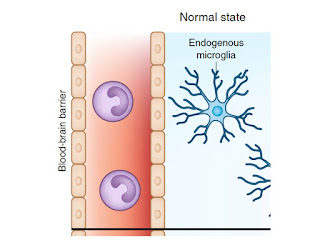
With the endothelium as its central unit, the   blood-brain barrier   (BBB) is a complex multicellular structure separating the central nervous system (CNS) from the systemic circulation. Disruption of the BBB has now been implicated in a multitude of acute and chronic CNS disorders indicating the potentially devastating effects of BBB breakdown on   brain   function. However, the healthy BBB is not an impermeable wall, but rather a communication 'centre', responding to and passing signals between the CNS and   blood . New studies are identifying BBB-specific transport pathways that tightly regulate the entry and exit of molecules to and from the   brain . They are revealing a highly plastic   barrier   in which dynamic changes in BBB components like paracellular tight junction complexes can contribute to BBB maintenance. Here, we provide a succinct overview of the current state-of-play in BBB research and summarize novel findings into BBB regulation in homeostatic r

The blood-brain barrier

Imagen
Blood   vessels are critical to deliver oxygen and nutrients to all of the tissues and organs throughout the body. The   blood   vessels that vascularize the central nervous system (CNS) possess unique properties, termed the   blood-brain barrier , which allow these vessels to tightly regulate the movement of ions, molecules, and cells between the   blood   and the   brain . This precise control of CNS homeostasis allows for proper neuronal function and also protects the neural tissue from toxins and pathogens, and alterations of these   barrier   properties are an important component of pathology and progression of different neurological diseases. The physiological   barrier   is coordinated by a series of physical, transport, and metabolic properties possessed by the endothelial cells (ECs) that form the walls of the   blood   vessels, and these properties are regulated by interactions with different vascular, immune, and neural cells. Understanding how these different cell

Microglia Development and function

Imagen
Proper development and function of the mammalian central nervous system (CNS) depend critically on the activity of parenchymal sentinels referred to as microglia. Although microglia were first described as ramified brain-resident phagocytes, research conducted over the past century has expanded considerably upon this narrow view and ascribed many functions to these dynamic CNS inhabitants. Microglia are now considered among the most versatile cells in the body, possessing the capacity to morphologically and functionally adapt to their ever-changing surroundings. Even in a resting state, the processes of microglia are highly dynamic and perpetually scan the CNS. Microglia are in fact vital participants in CNS homeostasis, and dysregulation of these sentinels can give rise to neurological disease. In this review, we discuss the exciting developments in our understanding of microglial biology, from their developmental origin to their participation in CNS homeostasis and pathophysiologic

Coxsackievirus Adenovirus Receptor Loss Impairs Adult Neurogenesis, Synapse Content, and Hippocampus Plasticity

Imagen
Although we are beginning to understand the late stage of neurodegenerative diseases, the molecular defects associated with the initiation of impaired cognition are poorly characterized. Here, we demonstrate that in the adult brain, the coxsackievirus and adenovirus receptor (CAR) is located on neuron projections, at the presynapse in mature neurons, and on the soma of immature neurons in the hippocampus. In a proinflammatory or diseased environment, CAR is lost from immature neurons in the hippocampus. Strikingly, in hippocampi of patients at early stages of late-onset Alzheimer’s disease (AD), CAR levels are significantly reduced. Similarly, in triple-transgenic AD mice, CAR levels in hippocampi are low and further reduced after systemic inflammation. Genetic deletion of CAR from the mouse brain triggers deficits in adult neurogenesis and synapse homeostasis that lead to impaired hippocampal plasticity and cognitive deficits. We propose that post-translational CAR loss of function

Physical biology of human brain development

Imagen
Neurodevelopment is a complex, dynamic process that involves a precisely orchestrated sequence of genetic, environmental, biochemical, and physical events. Developmental biology and genetics have shaped our understanding of the molecular and cellular mechanisms during neurodevelopment. Recent studies suggest that physical forces play a central role in translating these cellular mechanisms into the complex surface morphology of the human brain. However, the precise impact of neuronal differentiation, migration, and connection on the physical forces during cortical folding remains unknown. Here we review the cellular mechanisms of neurodevelopment with a view toward surface morphogenesis, pattern selection, and evolution of shape. We revisit cortical folding as the instability problem of constrained differential growth in a multi-layered system. To identify the contributing factors of differential growth, we map out the timeline of neurodevelopment in humans and highlight the cellular

HIV-associated synaptic degeneration

Imagen
Human immunodeficiency virus (HIV) infection induces neuronal injuries, with almost 50% of infected individuals developing HIV-associated neurocognitive disorders (HAND). Although highly activate antiretroviral therapy (HAART) has significantly reduced the incidence of severe dementia, the overall prevalence of HAND remains high. Synaptic degeneration is emerging as one of the most relevant neuropathologies associate with HAND. Previous studies have reported critical roles of viral proteins and inflammatory responses in this pathogenesis. Infected cells, including macrophages, microglia and astrocytes, may release viral proteins and other neurotoxins to stimulate neurons and cause excessive calcium influx, overproduction of free radicals and disruption of neurotransmitter hemostasis. The dysregulation of neural circuits likely leads to synaptic damage and loss. Identification of the specific mechanism of the synaptic degeneration may facilitate the development of effective therapeuti

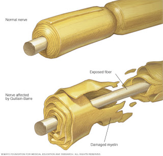
Guillain–Barré syndrome, transverse myelitis and infectious diseases

Imagen
Guillain–Barré syndrome (GBS) and transverse myelitis (TM) both represent immunologically mediated polyneuropathies of major clinical importance. Both are thought to have a genetic predisposition, but as of yet no specific genetic risk loci have been clearly defined. Both are considered autoimmune, but again the etiologies remain enigmatic. Both may be induced via molecular mimicry, particularly from infectious agents and vaccines, but clearly host factor and co-founding host responses will modulate disease susceptibility and natural history. GBS is an acute inflammatory immune-mediated polyradiculoneuropathy characterized by tingling, progressive weakness, autonomic dysfunction, and pain. Immune injury specifically takes place at the myelin sheath and related Schwann-cell components in acute inflammatory demyelinating polyneuropathy, whereas in acute motor axonal neuropathy membranes on the nerve axon (the axolemma) are the primary target for immune-related injury. Outbreaks of GBS

Complete genome sequence from Zika Virus, from Oaxaca, México

Imagen
Zika virus (ZIKV) is an emerging arthropod-borne flavivirus associated with severe congenital malformations and neurological complications. Although the ZIKV genome is well characterized, there is limited information regarding changes after cell isolation and culture adaptation. We isolated, and passaged in Vero cells, ZIKV from the serum of a symptomatic male patient and compared the viral genomes before and after culture. Single nucleotide polymorphisms were characteristic among serum-circulating genomes, while such diversity decreased after cell culture. REFERENCE: Boukadida, Celia et al. “ Complete Genome Sequences, before and after Mammalian Cell Culture, of Zika Virus Isolated from the Serum of a Symptomatic Male Patient from Oaxaca, Mexico. ” Genome Announcements 5.12 (2017): e00072–17. PMC. Web. 18 Jan. 2018.