Entradas

Mostrando las entradas de julio, 2018

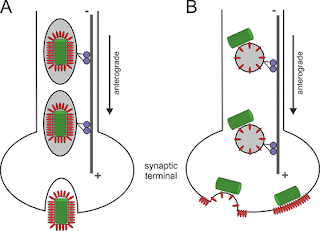
A "Driver Switchover" Mechanism of Influenza Virus Transport from Microfilaments to Microtubules.

Imagen
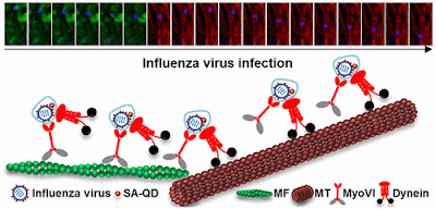
When infecting host cells, influenza virus must move on microfilaments (MFs) at the cell periphery and then move along microtubules (MTs) through the cytosol to reach the perinuclear region for genome release. But how viruses switch from the actin roadway to the microtubule highway remains obscure. To settle this issue, we systematically dissected the role of related motor proteins in the transport of influenza virus between cytoskeletal filaments in situ and in real-time using quantum dot (QD)-based single-virus tracking (SVT) and multicolor imaging. We found that the switch between MF- and MT-based retrograde motor proteins, myosin VI (myoVI) and dynein, was responsible for the seamless transport of viruses from MFs to MTs during their infection. After virus entry by endocytosis, both the two types of motor proteins are attached to virus-carrying vesicles. MyoVI drives the viruses on MFs with dynein on the virus-carrying vesicle hitchhiking. After role exchanges at actin-microtubul

Inhibition of Retrograde Transport Limits Polyomavirus Infection In Vivo

Imagen
Polyomaviruses (PyVs) silently infect most humans, but they can cause life-threatening diseases in immunocompromised individuals. The JC polyomavirus (JCPyV) induces progressive multifocal leukoencephalopathy, a severe demyelinating disease in multiple sclerosis patients receiving immunomodulatory therapy, and BK polyomavirus (BKPyV)-associated nephropathy is a major cause of kidney allograft failure. No effective anti-PyV agents are available. Several compounds have been reported to possess anti-PyV activity in vitro, but none have shown efficacy in clinical trials. Productive PyV infection involves usurping the cellular retrograde vesicular transport pathway to enable endocytosed virions to navigate to the endoplasmic reticulum where virion uncoating begins. Compounds inhibiting this pathway have been shown to reduce infection by simian virus 40 (SV40), JCPyV, and BKPyV in tissue culture. In this study, we investigated the potential of Retro-2.1, a retrograde transport inhibitor, t

Anterograde transport of rabies virus in DRG neurons.

Imagen
Model of intraneuronal postreplicative RABV transport. Rabies  virus  (RABV) spread is widely accepted to occur only by  retrograde  axonal  transport . However, examples of anterograde RABV spread in peripheral  neurons  such as dorsal root ganglion (DRG)  neurons  indicated a possible bidirectional  transport  by an uncharacterized mechanism. Here, we analyzed the axonal  transport  of fluorescence-labeled RABV in DRG  neurons  by live-cell microscopy. Both entry-related  retrograde   transport  of RABV after infection at axon endings and postreplicative  transport  of newly formed  virus  were visualized in compartmentalized DRG neuron cultures. Whereas entry-related  transport  at 1.5 μm/s occurred only retrogradely, after 2 days of infection, multiple particles were observed in axons moving in both the anterograde and  retrograde  directions. The dynamics of postreplicative  retrograde   transport  (1.6 μm/s) were similar to those of entry-related  retrograde   transport . In

Flaviviruses are neurotropic, but how do they invade the CNS?

Imagen
Flaviruses (FV) are RNA viruses carried by mosquitoes. Neurological signs including acute encephalitis, meningitis and acute flaccid paralysis develop in a small percentage of infected individuals; long term sequlae are, Parkinsonism, dystonias and cognitive changes. FV neuroinfection is neurotropic involving subcortical nuclei (substantia nigra and thalamus) anterior horn neurons and neocortex. Glycosylation of the FV E envelope protein is one determinant of neuroinvasion, increasing both axonal and trans-epithelial transportation. Neutralizing antibodies against the E and NS proteins prevents FV uptake into several cell types, including axons. CD8+ T cells are vital for clearance of WNF infected cells from the CNS, whereas TLR-3 and TLR-7 mediated anti-virus response through increased serum inflammatory cytokines to disrupt the BBB providing infected leucocytes and free virus access to the CNS (so called Trojan horse) Cellular virus attachment factors, promoting FV cell entry are w

Prions, prionoids and protein misfolding disorders #PMD

Imagen
Prion diseases are progressive, incurable and fatal neurodegenerative conditions. The term ‘prion’ was first nominated to express the revolutionary concept that a protein could be infectious. We now know that prions consist of PrPSc, the pathological aggregated form of the cellular prion protein PrPC. Over the years, the term has been semantically broadened to describe aggregates irrespective of their infectivity, and the prion concept is now being applied, perhaps overenthusiastically, to all neurodegenerative diseases that involve protein aggregation. Indeed, recent studies suggest that prion diseases (PrDs) and protein misfolding disorders (PMDs) share some common disease mechanisms, which could have implications for potential treatments. Nevertheless, the transmissibility of bona fide prions is unique, and PrDs should be considered as distinct from other PMDs. REFERENCE: Scheckel C, Aguzzi A. Prions, prionoids and protein misfolding disorders. Nat Rev Genet. 2018 Jul;19(7):

Clinical Trials in Neurovirology: Successes, Challenges, and Pitfalls

Imagen
Clinical trials in neurovirology illustrate the special challenges confronting investigators planning to study these conditions, as well as the contributions of successful trials in establishing appropriate management for these devastating diseases. This article reviews key examples of progress in neurovirology that have been spurred by clinical trials, emphasizing human herpes virus encephalitis, HIV, and JC virus. Clinical trials in the setting of neurovirological diseases are characterized by specific challenges, which may include small sample sizes, clinical presentations from life-threatening conditions to chronic courses of disease, regional and temporally restricted outbreaks scenarios, and the unavailability of validated diagnostic tests that can be rapidly deployed at the bedside. This review aims to highlight these methodological challenges and pitfalls in designing and executing clinical neurovirology trials, as well as to outline innovative trial designs, which could be

#brain: The mysterious origins of microglia

Imagen
The origin of microglia, the resident macrophage population of the CNS, has been a long-standing matter of debate. Here we discuss two seminal studies published in 2007 in Nature Neuroscience that significantly contributed to a better understanding of microglia ontogeny and homeostasis in the adult brain. REFERENCE: Ginhoux F, Garel S. The mysterious origins of microglia. Nat. Neurosci. 2018 Jul;21(7):897-899. doi: 10.1038/s41593-018-0176-3.

Viral Triggers and Inflammatory Mechanisms in Pediatric Epilepsy. Review

Imagen
Experimental and clinical findings suggest a crucial role for inflammation in the onset of pediatric seizures; this mechanism is not targeted by conventional antiepileptic drugs and may contribute to refractory epilepsy. Several triggers, including infection with neurotropic viruses such as human herpesvirus 6 (HHV-6), other herpesviruses, and picornaviruses, appear to induce activation of the innate and adaptive immune systems, which results in several neuroinflammatory responses, leading to enhanced neuronal excitability, and ultimately contributing to epileptogenesis. This review discusses the proposed mechanisms by which infection with herpesviruses, and particularly with HHV-6, and ensuing inflammation may lead to seizure generation, and later development of epilepsy. We also examine the evidence that links herpesvirus and picornavirus infections with acute seizures and chronic forms of epilepsy. Understanding the mechanisms by which specific viruses may trigger a cascade of alt

Differential antiviral immunity to Japanese encephalitis virus in developing cortical organoids

Imagen
Japanese encephalitis (JE) caused by Japanese encephalitis virus (JEV) poses a serious threat to the world's public health yet without a cure. Certain JEV-infected neural cells express a subset of previously identified intrinsic antiviral interferon stimulated genes (ISGs), indicating brain cells retain autonomous antiviral immunity. However, whether this happens in composited brain remains unclear. Human pluripotent stem cell (hPSC)-derived organoids can model disorders caused by human endemic pathogens such as Zika virus , which may potentially address this question and facilitate the discovery of a cure for JE. We thus generated telencephalon organoid and infected them with JEV. We found JEV infection caused significant decline of cell proliferation and increase of cell death in brain organoid, resulting in smaller organoid spheres. JEV tended to infect astrocytes and neural progenitors, especially the population representing outer radial glial cells (oRGCs)

Virus-like particles and enterovirus antigen found in the brainstem neurons of #Parkinson's disease

Imagen
Background: In a previous study on encephalitis lethargica, we identified a virus related to enterovirus in autopsy brain material. Transmission electron microscopy (TEM), immunohistochemistry (IHC) and molecular analysis were employed.  Our present objective was to investigate, using a similar approach, as to whether virus -like particles (VLP) and enterovirus antigen are present in Parkinson's disease (PD) brainstem neurons. Methods: Fixed tissue from autopsy specimens of late onset PD and control brainstem tissue were received for study. The brain tissue was processed for TEM and IHC according to previous published methods. Results:  We observed VLP in the brainstem neurons of all the cases of PD that were examined.  In the neurons' cytoplasm there were many virus factories consisting of VLP and endoplasmic reticulum membranes. In some neurons, the virus factories contained incomplete VLP. Complete VLP in some neurons' virus factories had an average diam