Entradas

Estimating the prevalence at death of CTE neuropathology among professional football players

Imagen
In July 2017,   JAMA   published results from autopsies of the brains of 202 deceased American football players. 1   Of the 111 participants who had played in the National Football League (NFL), 110 (99%) received a neuropathologic diagnosis of chronic traumatic encephalopathy (CTE), a progressive neurodegenerative disease associated with head trauma. This 99% figure received substantial media coverage when the study was published. REFERENCE; Binney ZO, Bachynski KE. Estimating the prevalence at death of CTE  neuropathology among professional football players. Neurology. 2018 Nov 28. pii:   10.1212/WNL.0000000000006699. doi: 10.1212/WNL.0000000000006699. [Epub ahead of  print] PubMed PMID: 30487144. Commented on: InsideScience: What's the Risk of Chronic Traumatic Encephalopathy for NFL Players? New study suggests at least one in 10 NFL players could eventually develop the brain disease.

TWiV 453: Neurovirology with Diane Griffin

From the Vector-Borne Viruses Symposium in Hamilton, Montana, Dickson and Vincent speak with Diane Griffin about her career and her work on understanding viral infections of the central nervous system. Hosts: Vincent Racaniello and Dickson Despommier Guest: Diane Griffin TWiV 453 (39 MB .mp3, 64 min) LINKS FOR THIS EPISODE Vector-Borne Viruses Symposium Diane Griffin Adoptive immunization of immunosuppressed mice infected with Sindbis virus Role of the immune response in age-dependent resistance of mice to encephalitis due to Sindbis virus Antibody-mediated clearance of alphavirus infection from neurons Persistence of viral RNA in mouse brains after recovery from acute alphavirus encephalitis Conversion of lytic to persistent alphavirus infection by the bcl-2 cellular oncogene Bc1-2 protects mice against fatal alphavirus encephalitis

8º Curso de Animales de Laboratorio #INR 12-14/SEP/2018

 ·

A "Driver Switchover" Mechanism of Influenza Virus Transport from Microfilaments to Microtubules.

Imagen
When infecting host cells, influenza virus must move on microfilaments (MFs) at the cell periphery and then move along microtubules (MTs) through the cytosol to reach the perinuclear region for genome release. But how viruses switch from the actin roadway to the microtubule highway remains obscure. To settle this issue, we systematically dissected the role of related motor proteins in the transport of influenza virus between cytoskeletal filaments in situ and in real-time using quantum dot (QD)-based single-virus tracking (SVT) and multicolor imaging. We found that the switch between MF- and MT-based retrograde motor proteins, myosin VI (myoVI) and dynein, was responsible for the seamless transport of viruses from MFs to MTs during their infection. After virus entry by endocytosis, both the two types of motor proteins are attached to virus-carrying vesicles. MyoVI drives the viruses on MFs with dynein on the virus-carrying vesicle hitchhiking. After role exchanges at actin-microtubul...

Inhibition of Retrograde Transport Limits Polyomavirus Infection In Vivo

Imagen
Polyomaviruses (PyVs) silently infect most humans, but they can cause life-threatening diseases in immunocompromised individuals. The JC polyomavirus (JCPyV) induces progressive multifocal leukoencephalopathy, a severe demyelinating disease in multiple sclerosis patients receiving immunomodulatory therapy, and BK polyomavirus (BKPyV)-associated nephropathy is a major cause of kidney allograft failure. No effective anti-PyV agents are available. Several compounds have been reported to possess anti-PyV activity in vitro, but none have shown efficacy in clinical trials. Productive PyV infection involves usurping the cellular retrograde vesicular transport pathway to enable endocytosed virions to navigate to the endoplasmic reticulum where virion uncoating begins. Compounds inhibiting this pathway have been shown to reduce infection by simian virus 40 (SV40), JCPyV, and BKPyV in tissue culture. In this study, we investigated the potential of Retro-2.1, a retrograde transport inhibitor, t...

Anterograde transport of rabies virus in DRG neurons.

Imagen
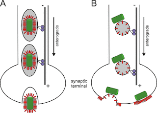
Model of intraneuronal postreplicative RABV transport. Rabies  virus  (RABV) spread is widely accepted to occur only by  retrograde  axonal  transport . However, examples of anterograde RABV spread in peripheral  neurons  such as dorsal root ganglion (DRG)  neurons  indicated a possible bidirectional  transport  by an uncharacterized mechanism. Here, we analyzed the axonal  transport  of fluorescence-labeled RABV in DRG  neurons  by live-cell microscopy. Both entry-related  retrograde   transport  of RABV after infection at axon endings and postreplicative  transport  of newly formed  virus  were visualized in compartmentalized DRG neuron cultures. Whereas entry-related  transport  at 1.5 μm/s occurred only retrogradely, after 2 days of infection, multiple particles were observed in axons moving in both the anterograde and  retrograde  directions. The dy...

Flaviviruses are neurotropic, but how do they invade the CNS?

Imagen
Flaviruses (FV) are RNA viruses carried by mosquitoes. Neurological signs including acute encephalitis, meningitis and acute flaccid paralysis develop in a small percentage of infected individuals; long term sequlae are, Parkinsonism, dystonias and cognitive changes. FV neuroinfection is neurotropic involving subcortical nuclei (substantia nigra and thalamus) anterior horn neurons and neocortex. Glycosylation of the FV E envelope protein is one determinant of neuroinvasion, increasing both axonal and trans-epithelial transportation. Neutralizing antibodies against the E and NS proteins prevents FV uptake into several cell types, including axons. CD8+ T cells are vital for clearance of WNF infected cells from the CNS, whereas TLR-3 and TLR-7 mediated anti-virus response through increased serum inflammatory cytokines to disrupt the BBB providing infected leucocytes and free virus access to the CNS (so called Trojan horse) Cellular virus attachment factors, promoting FV cell entry are w...